Dangerous Decibels Program Walk-Through: 3. How Do We Hear?
“How Do We Hear?” Recap
Reminders
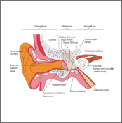
- Teach the following anatomical parts:
- Pinna
- Eardrum
- 3 small bones in the middle ear
- Cochlea
- Hair Cells and Hair Bundles
- Hearing nerve
- Brain
- Explain how sound vibrations are transmitted through the parts of the ear.
- Emphasize that the cochlea is filled with thousands of tiny sensors called “hair cells” and on top of the hair cells are even smaller hair like structures call “hair bundles”.
- When “hair bundles” rock back and forth, that creates a signal to the rest of the hair cell which in turn send a signal through the hearing nerve to the brain.
- It is our brain that can identify the sound.
Key Points
Materials
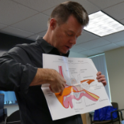
- It is a good idea to have the ear anatomy poster laminated so that it lasts longer.
- Make sure the poster is visible to everyone in the classroom.
- You are provided an electronic version of the ear anatomy poster that you can download under RESOURCES. This will enable you to print replacement copies or print even larger posters to use in the classroom if desired. Larger posters will be more visible to students, but harder for the educator to hold and point to.
- In some instances, the educator will use a computer to project the ear anatomy poster in the classroom so that it is larger and easier to read. If this is the approach taken, make sure you have a pointer (or electronic pointer) to differentiate the parts of the ear when teaching “How Do We Hear”.